Magnetic Resonance and Bone Changes at the Enthesis
Magnetic Resonance Imaging of the Enthesis in patients with Swollen Joints
Patients with Enthesis related arthritis may come to medical attention with knee joint swelling.
Due to pain and swelling and distension of the joint the doctor may not be able to appreciate inflammation at insertions or enthesitis.
When patients go to see an Orthopedic surgeon with a swollen knee they may have a general anesthetic and have a camera inserted into the swollen joint (arthroscopy).
However, the diseased insertions are hidden underneath the joint lining and no abnormality will be seen and the diagnosis will be missed.
If a conventional Magnetic Resonance Imaging scan is requested then the inflammation at the enthesis and bone may also be missed as shown below.
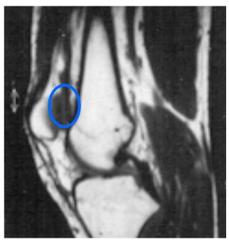 |
This is a Magnetic Resonance Imaging scan from a young man with psoriasis and knee swelling. The only abnormality is free fluid in the joint or a joint effusion (inside blue circle). This image was obtained using what is called a T1-weighted image. In this picture the gray area is fat within the bone marrow. |
A more specialized type of Magnetic Resonance Imaging scan called fat suppression MRI can pick up the enthesis related pathology.
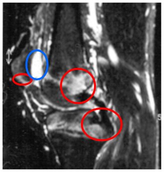 |
This is the same patient who now has had a different type of Magnetic Resonance Imaging picture obtained in the same scanner immediately after the image above. The free joint fluid now appears white (inside blue circle). In this image the signal from the fat is suppressed and now inflammation can be clearly seen at 3 insertion regions (inside red circles). This is enthesitis at multiple sites which is not evident to the doctor on clinical examination. This type of scan is called a fat suppressed scan. |
So in subjects with a swollen knee or other joint, fat suppression Magnetic Resonance Imaging can be used to accurately diagnose the enthesitis related arthritis.
Problems and limitations of MRI for enthesitis diagnosis
The doctors that look at MRI scans that are not knowledgeable on the importance of the enthesis may also miss subtle diagnostic changes.
Other changes including new bone formation at insertions may be missed.
Although MRI is good, it is not perfect. Because insertions are difficult to see on MRI the scan may be totally normal in cases with enthesitis related pain.
Ultrasound is an alternative to evaluate such cases.
The use of Magnetic Resonance Imaging for the diagnosis of Enthesitis is an ongoing area of research.
References
