MRI of Damage in Normal Insertions
Introduction
One might think that people without joint symptoms would have normal MRI scans of their entheses and that only patients with Ankylosing Spondylitis (AS) and Psoriatic Arthritis (PsA) and the allied with Spondyloarthropathies would have abnormal scans. This page explains why normal people without any arthritis may have abnormalities on MRI scanning of the enthesis. This is part of the normal cycle of repair at these sites that are prone to microdamage.
Mechanism
The entheses is a site of high mechanical stressing and reflecting this normal insertions may get damaged and undergo repair. The levels of damage related changes visible on MRI in normal may be identical to those seen in subjects with PsA and AS and other diseases including osteoarthritis. Some examples of these changes in normal people are shown below.
Normal Sacroiliac Joint bone damage
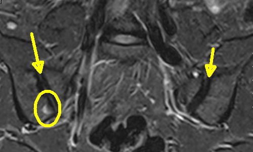
What is termed as "bone oedema" (bone edema {US}) meaning increased water content due to blood vessel leakiness is not uncommon in the sacroiliac joint when viewed using MRI. These changes can be induced by physical activity, are painless and resolve within 6 weeks [1]. The changes in the sacroiliac joints in normal people tend to occur at the joint capsule enthesis to bone or at he joint margin.
|
| this is a MR image of the sacroiliac joint. The yellow arrows show the joint space. Inside the yellow circle is high signal (white) which is termed a patch of bone oedema. This pattern is evident in some normals but also in AS. |
Normal spine bone damage
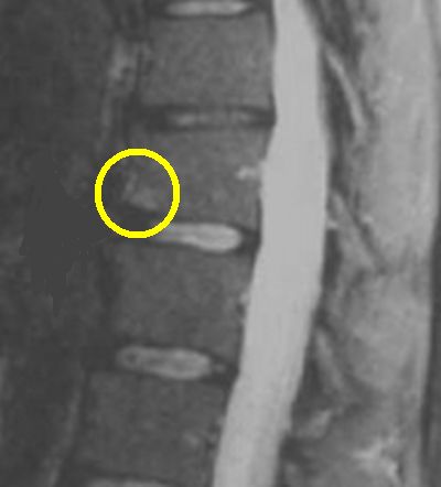
Spinal entheses bone oedema reactions on MRI are seen in completely normal subjects. It is likely that the HLA-B27 gene and gut leakiness determine if such subtle harmless abnormalities may progress [2].
|
| this is a MR image of the spine. Inside the yellow circle is a patch of bone oedema that looks identical to the pattern seen in AS and Spondyloarthropathy. We believe that these abnormalities are related to the normal repair processes that take place as a result of the high stressing exerted on spinal entheses. |
Other sites of damage
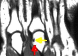
The attachment sites form enthesis organs and enthesis tissue compresses the adjacent bone. This can lead to microscopic erosions that are not visible on X-ray but are seen on MRI.
|
| this is a MR image of normal knuckles (MCP) joints. The red arrow shows the site of the capsule enthesis. The enthesis organ compresses the adjacent bone. In this case the associated stressing has lead to a microscopic bone erosion. |
Strategy for dealing with these findings
A good knowledge of the enthesis and its response to stress should stop doctors putting too much emphasis on MR scanning alone. A careful patient history is key to avoid a wrong diagnosis.
Repeat MR scanning could be considered if any doubt exists about the diagnosis. In this setting changes should resolve and new ones should be unlikely to develop.
Multiple entheseal related abnormalities or bigger regions of abnormality are more likely to be linked to disease rather than normal repair reactions.
Normal level stressing related changes should clear up within a few months. If the entheis lesion is beside a damaged tissue structure such as a disc changes may persist.
Implications
There is a clear danger that a lack of knowledge about the biology of the enthesis, even amongst people who are considered experts, might lead to a wrong diagnosis of AS, PsA or axial Spondyloarthritis. An abnormal MRI scan does not necessarily mean AS, PsA or SpA.
This may lead to prescription of biological drugs which are costly, risk side effects and won't work in this wrong setting.
References

Resources
MRI finds runners' overuse injuries. Overuse injuries have a tendency to affect the same territories as Spondyloarthropathy including the lower limbs.