Predicting AS in young people with back pain
Introduction
Historically it took up to a decade, or longer for a diagnosis of Ankylosing Spondylitis (AS) to be made. The reason for this was that the diagnostic changes evident on sacroiliac joint X-rays were slow to develop. This page explains how patients destined to develop AS can now be recognised in the early stages of disease.
Recognition of AS prior to X-ray changes
The advent of fat suppression MRI has transformed the early recognition of AS. Studies have shown that MRI defined "bone oedema" that is due to bone marrow inflammation (osteitis) beneath the sacroiliac joint fibrocartilage can be used to predict who will get AS in the following decade. A series of MRI images are shown that relate to the development of AS.
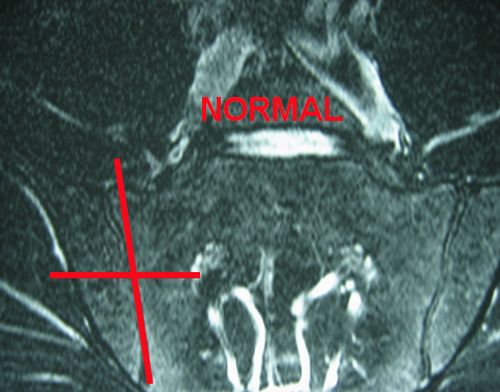
Normal MRI scan of sacroiliac joint
This is a normal image of the sacroiliac joint. Patients with early inflammatory back pain that may go on to develop AS could on occasion have a normal scan because the inflammation comes and goes.
|
| This is a normal MRI. The read grid is the Leeds based scoring system to quantify the bone oedema |
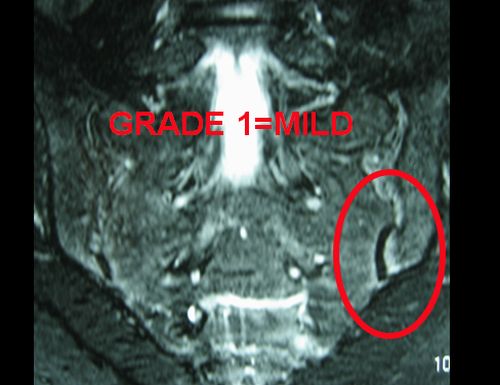
Mild MRI determined Bone Oedema
A small focus of bone oedema on MRI is not that strong a predictor for the development of AS. Such changes may be seen in normal subjects or after vigorous activity including exercise.
|
| This shows severe oedema. The likelihood of this progressing is low. |
There is a danger that these harmless changes of potentially no relevance are being used to make a diagnosis of "axial Spondyloarthritis" leading to the use of biological therapies.
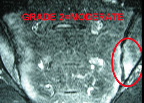
Moderate Bone Oedema
This has very predictive for the development of AS and especially so in HLA-B27 positive cases. This is because HLA-B27 is a factor that predicts recurrent or chronic bone oedema in the sacroiliac joint and spine.
|
| A moderate degree of bone oedema is shown inside the red circle. This is quite likely to progress to AS and more so if the patient with back pain is HLA-B27 positive. |
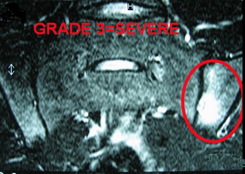
Severe Bone Oedema
The likelihood of developing AS is greatly increased if there is extensive sacroiliac joint bone oedema. In fact cases were found to be 13 times more likely to get AS if severe bone oedema at baseline.
|
| The yellow arrows show the diffuse osteitis (white) in both sides of the sacroiliac joint |
Implications
These cases of MRI positive disease are currently called axial Spondylarthritis reflecting the fact the x-ray changes are not evident. However, such cases have the same degree of disability as patients with early AS and positive X-rays. This is an area of intensive research to define severe early disease.
References
